Hysterosalpingography (HSG) :-
What is hysterosalpingography (HSG)?
Hysterosalpingography (HSG) is an X-ray procedure that is used to view the inside of the uterus and fallopian tubes. It often is used to see if the fallopian tubes are partly or fully blocked. It also can show if the inside of the uterus is a normal size and shape.
Why is HSG done?
- Scarring or abnormalities in the uterus or fallopian tubes can lead to infertility and pregnancy problems.
- HSG also is used a few months after some tubal sterilization procedures to make sure that the fallopian tubes have been completely blocked.
When is HSG not done?
- HSG is not done if you
- are pregnant
- have a pelvic infection
- have uterine bleeding at the time of the procedure
What should I do to prepare for HSG?
- Your obstetrician–gynecologist (ob-gyn) may recommend that you take an over-the-counter pain reliever an hour before the procedure. Discuss this decision with your ob-gyn. In some cases, you also may receive an antibiotic for you to take before HSG.
- Most people can drive themselves home after having HSG. But you may not feel well after the procedure, so you may want to make arrangements for someone to drive you home.
How is HSG done?
- HSG is done in a hospital, clinic, or the office of your ob-gyn. It is best to have HSG done in the first half of the menstrual cycle (days 1 to 14). This timing reduces the chance that you may be pregnant.
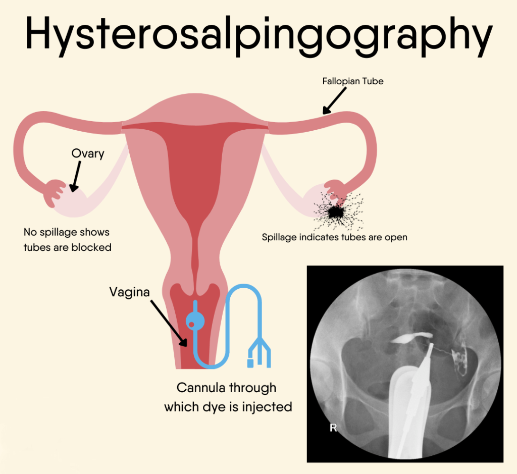
- During HSG, a contrast agent is placed in the uterus and fallopian tubes. This is a fluid that contains a dye. The dye shows up in contrast to the body structures on an X-ray screen. The dye outlines the inner size and shape of the uterus and fallopian tubes. It also is possible to see how the dye moves through the body structures.
- The procedure is done in the following way:
- You lie on your back with your feet placed as for a pelvic exam. A device called a speculum is inserted into the vagina. It holds the walls of the vagina apart to allow the cervix to be viewed. The cervix is cleaned.
- The end of the cervix may be injected with local anesthesia (pain relief). You may feel a slight pinch or tug as this is done.
- One of two methods may be used to insert the dye. In one method, the cervix is grasped with a device to hold it steady. An instrument called a cannula is then inserted into the cervix. In the other method, a thin plastic tube is passed into the cervical opening. The tube has a small balloon at the end that is inflated. The balloon keeps the tube in place in the uterus.
- The speculum is removed, and you are placed beneath an X-ray machine. The fluid is placed through the cannula or tube into the uterus and fallopian tubes. The fluid may cause cramping. If the tubes are blocked, the fluid will cause them to stretch.
- X-ray images are taken as the fluid fills the uterus and tubes. You may be asked to change position. If there is no blockage, the fluid will spill slowly out the far ends of the tubes. After it spills out, the fluid is absorbed by the body.
- After the images are taken, the speculum and cannula or tube is removed.
What should I expect after the procedure?
- After HSG, you can expect to have sticky vaginal discharge as some of the fluid drains out of the uterus. The fluid may be tinged with blood. A pad can be used for the vaginal discharge. Do not use a tampon. You also may have the following symptoms:
- Slight vaginal bleeding
- Cramps
- Feeling dizzy, faint, or sick to your stomach