
LEVEL 2 ULTRASOUND
A level II ultrasound is similar to a standard ultrasound. The difference is that your doctor will get more detailed information. Your doctor may focus on specific parts of your baby’s body, such as their brain, heart, or the other organs.
You may get a targeted ultrasound in your second trimester. It can help check your baby for some birth defects, such as Down syndrome.
How the Test Is Done
- A level II ultrasound is just like a regular abdominal ultrasound. You’ll lie down and a technician will put a special gel on your belly. This will help carry the sound waves. Then the technician will hold a probe against your belly and move it around to get an image. You may need to go into the test with a full bladder. This will make the test results more clear.
What to Know About Test Results
- Your doctor will probably give you the results after the exam. A normal result should be reassuring. However, keep in mind that ultrasounds are not always accurate. They can’t diagnose or rule out many problems.
What to Expect During a Level 2 Ultrasound
This ultrasound (or anatomy scan) is done
about halfway through pregnancy
A detailed anatomy ultrasound is recommended during pregnancy, between 18 and 22 weeks gestation.1 Also known as a level 2 ultrasound, anatomy scan, or anomaly scan, the aim of this imaging is for your provider to assess your baby’s health and development.
While the earlier, level 1 ultrasounds look to measure the baby and note its position, a level 2 ultrasound looks in greater detail at the different parts of your baby’s body. Generally, they will examine the baby’s whole body, including their brain, heart, and other organs. The umbilical cord and amniotic fluid levels will also be checked.
This is typically a transabdominal ultrasound, as opposed to a transvaginal scan like those done earlier in pregnancy. This test is unlike any other performed in early pregnancy, and can be a nerve-wracking experience for expectant parents. But with a little bit of knowledge and preparation, it doesn’t have to be.
What Happens at a Level 2 Ultrasound?

To begin the scan, the ultrasound technician (or in some cases your doctor) will have you lie down on the exam table. Next, an ultrasonic gel (which is warmed for your comfort) will be placed on your belly.
The technician or your physician will move the ultrasound transducer wand over your abdomen. You will feel them pressing on your belly, but it won’t hurt you or your baby. However, if you do feel uncomfortable at any time during the scan, it’s important to let your provider know.
During the ultrasound, fetal anatomy and size are assessed. The entire anomaly scan typically takes between 45 and 75 minutes from start to finish, and you often receive results with the same appointment. The size of your baby in comparison to other babies of the same gestational age, as well as the placenta, will be evaluated to ensure your baby is developing properly.
What a Level 2 Ultrasound Looks At
Between 18 and 22 weeks into your pregnancy, your baby will be large enough for your medical provider to see details of their organs and limbs from head to toe. If you’re having multiples, each fetus will receive their own scan.
Sometimes, follow-up ultrasounds for growth and reevaluation will be suggested based on the findings of the initial scan. Barring any specific high-risk issues, the following areas are examined to rule out birth defects and other anomalies or red flags:
- Brain (including ventricles, cerebellum, corpus callosum, and other key structures)
- Neck (including nuchal fold thickness)
- Face structures (palate, eyes, nose, lips, and ears)
- Heart and lungs
- Spine and ribs
- Abdominal organs (stomach intestines, spleen, liver, gall bladder) and the abdominal wall
- Limbs and digits
- Genitals (if visible, can determine fetal sex)
- Umbilical cord, including vessels and insertion site
- Placenta structure and location
- Cervical length
- Fetal position and movements
- Amniotic fluid
Typically, the images produced by the ultrasound will appear on a screen where you and your provider or technician can see them. The images will be measured and recorded, and you may be given some printouts or a disc with the images to have as keepsakes.
Some hospitals or clinics will let you record the ultrasound session with a recording device, such as your cell phone camera. However, the rules around this vary from doctor to doctor. It’s best to ask about specific guidelines before your appointment.
Week 19 of Your Pregnancy

- Moments after birth, the first thing many new parents do is count 10 tiny fingers and 10 tiny toes. However, ultrasound technology enables providers and parents alike to count fingers and toes before a baby is actually born.1
- The potential of seeing the details like the fingers and toes of your baby during the scan can be something that can offer some joy and peace of mind if you’re feeling anxious about your scan.
- However, actually being able to count each finger and toe in an ultrasound exam depends on how cooperative your baby is during the scan. If they’re moving too much, it’s possible that you may still have to wait until birth to count all fingers and toes individually.
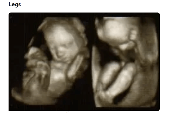
- Unlike the fingers and toes, it is very likely that you will be able to clearly see bigger areas like your baby’s arms and legs on an ultrasound despite their movement.
- During the scan, the ultrasound technician will measure your baby’s thigh bone (femur). This measurement helps them calculate how well your baby is growing for their gestational age.1
- Sometimes it’s also possible to see the baby kicking, which you may or may not feel at this point in your pregnancy—another potentially exciting moment to look forward to.
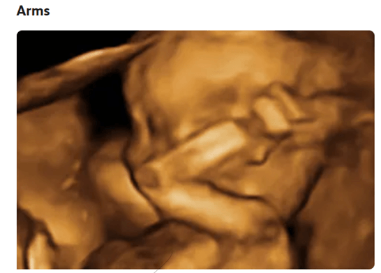
- In addition to looking at your baby’s legs, the ultrasound technician will also measure the bones of the arms (radius, ulna). Depending on their position, you may see your baby waving their arms or sucking their thumb.1
- Some technicians may even be able to snap a still photo of your baby’s arm waving or their thumb firmly planted in their mouth. This type of photo can become a cherished keepsake and way to remember the moment.

During the scan, the ultrasound technician will check your baby’s internal structures, including their brain.
At this point in fetal development, the appearance of the brain often has a butterfly look. The ultrasound technician will be examining for the presence and anatomic landmarks of your baby’s brain. They will also note any anomalies found, such as choroid plexus cysts.
The ultrasound technician will also look at your baby’s stomach, urinary tract, kidneys, and other structures.2 While every provider is different, you can typically expect to be informed of any unusual findings or abnormalities before the conclusion of your appointment.
Although the potential of finding abnormalities within your baby’s organs can be concerning, your provider will go over next steps with you if this occurs. If any problems are found, you will likely have further ultrasound exams that are usually conducted by specialists called perinatologista or maternal-fetal experts.
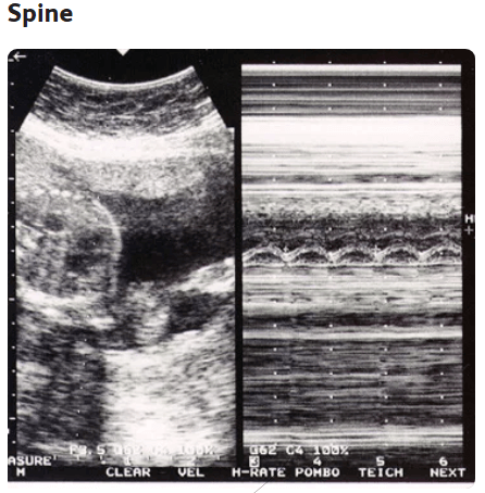
- Your baby’s spine will also be examined during the fetal anatomy survey.2
- This is usually one of the most easily recognizable anatomical elements for parents as they watch the ultrasound.
- The technician will confirm that your baby’s spine and neural tubeare completely formed and without cysts. Once again, if abnormalities are present, your provider will go over these issues with you—usually before the end of your appointment.

- During the ultrasound, the technician will look through your baby’s heart to measure the fetal heart rateand look for any structural problems, such as defects in the ventricles and other heart anomalies.2
- If they suspect a problem with your baby’s heart, they will most likely recommend a fetal echocardiogram.
- It’s important to remember that a suspected issue found in an anatomy scan does not automatically mean that a problem actually exists. Sometimes additional tests reveal that the suspected problem is either easily remedied, will be outgrown, or was just an abnormality that showed up in the initial scan, but is not concerning.
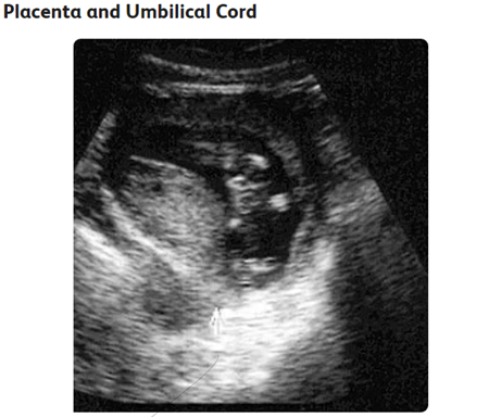
- During your anatomy scan, the technician will also closely examine the placenta.4Confirming where the placenta is located helps the technician screen you for a condition called placenta previa.
- An ultrasound exam also provides a good view of the umbilical cord to confirm that the cord functions properly. A normal umbilical cord has two arteries and one vein. The technician will check to see if you have a three-vessel or two-vessel cord (single umbilical artery).1
- Babies born with a two-vessel cord sometimes experience growth issues and other potential anatomic anomalies, so it’s important to be aware of the cord’s structure. However, most babies born with a two-vessel cord will have a normal fetal echocardiogram and be born perfectly healthy.